Pelotas, Sao Paulo E Brasilia,
Urgente, Newswire
A presidente da associacao de blogueiros de vinho de Capao Redondo publicou ontem uma carta aberta em todos os grandes jornais do pais denunciando uma guerra psicologica que tem como intuito abalar a credibilidade, vendas, producao e toda cadeia do vinho de uva no pais.
Segundo a presidente da associacao Paula Notobas, ha uma campanha promovida por agentes terroristas de mercado que tentam sacar beneficios as custas do liquido precioso e do medo do publico em geral. Essa rede de agentes espalha boatos infundados como ''vinho espumante gaucho nao he o segundo melhor do mundo'', ou ''ha muito vinho importado que custa muito, mas muito mais que vale'', ''ha vinhos que mal contem uva dentro da garrafa'', ''muito vinho argentino foi comprado no chile para ser engarrafado na argentina'' ou ainda a horrivel calunia ''as promocoes de internet sao armadilhas que funcionam sozinhas, parece que as pessoas querem pular dentro da jaula, nos nem esperavamos que fossem tao trouxas''.
A associacao de blogueiros de vinho de Capao Redondo esta organizando um protesto para o dia 31 de dezembro na avenida paulista, perto da meia noite. Espera-se que milhares de pessoas atendam ao clamor.
Monday, December 30, 2013
Sunday, December 29, 2013
Perguntar Ofende: Schumacher, Deus E O Bambu
Escrevo essa sem saber o desfecho da pancada que o Schumy (nos numeros o maior de todos, no talento que deu resultado tambem entre os top 5) deu nos alpes franceses, mas nao pude deixar de perceber que varias pessoas enviaram mensagem via internet dizendo que estao rezando para deus protege-lo (Schumacher).
Me pergunto se houvesse um deus por que ele/ela deixaria isso acontecer para comeco de conversa? Faz sentido deixar a desgraca acontecer para depois ''proteger'' o coitado apos reza/pedido dos fieis?
Faz sentido rezar para "deus" curar alguem com doenca terminal se "deus" -para comeco de conversa- poderia ter deixado a pessoa sem passar por essa?
Maximo que se pode fazer he torcer pela recuperacao da pessoa, sabendo que ''ondas positivas'' ou reza nao tem efeito algum tambem para essas coisas. Estatisticamente comprovado; sinto, turma da religiao.
Ah, queimar incenso prejudica vias areas e pular onda na praia dia 31 da doenca.
Tomara que o Schumy saia dessa ileso.
Saturday, December 28, 2013
Brazil X USA: Diferenca Abismal. Leitura De Sabado
Nesse feriado maravilhoso (25/12), pude colocar parte das leituras em dia. Uma em particular me chamou bem a atencao.
Mais uma diferenca gritante entre 'nos' e 'eles'. Dessa vez no uso da informacao. Apos ler o artigo (link AQUI http://www.economist.com/news/united-states/21591855-novel-way-measure-influence-protest-movement-watery-tea) , gostaria que voce pensasse se apos toda a onda de protesto com black bibas alguem coletou algum dado e se sim, se fez uso relevante dele. USA usa estatistica para tudo, e essa tal de estatistica preve muita coisa, de comportamento de germes a mercados acionarios.
Aqui estatistica he aquela coisa que aparece em futebol na globo, numero de passes e desarmes por jogador ou passes errados (no futebol americano funciona, no soccer, nao)
Boa leitura e reflexoes. Leitura curta.
Friday, December 27, 2013
Musica De Sexta-Feira
Jesus, ala, moises...quem trabalha nao percebe o espaco-tempo passando.
Ultima sexta do 'ano'. Mientras tanto un Iggy Pop para alegrar a vida.....
Thursday, December 26, 2013
Mais Que Uma Perola, Mas Uma Joia.....
http://classificados.folha.uol.com.br/empregos/2013/12/1390190-mcdonalds-pede-que-funcionarios-evitem-fast-food.shtml
Hilario. Mesma coisa que a Ford dizer que os funcionarios nao devem comprar ou dirigir os carros deles porque sao inseguros, a PMorris proibir funcionarios de fumar, importador de vinho contar aos funcionarios que os vinhos deles sao podres, academia de ginastica proibir funcionarios de malhar porque os aparelhos sao piratas e perigosos, a sococo dizer que agua de coco da cancer...e por ai vai.
Piada real para terminar bem a semana.
Tuesday, December 24, 2013
Qual O Vinho Do Ano De 2013 Para Voce?
Alguem ai teve um vinho que marcou o ano de alguma maneira, boa ou ruim?? Qual o criterio para tal escolha?
Foi o ano que mais comprei e vendi vinho, e ainda assim bebi pouquissimo. Degustacoes sim, mas garrafas poucas.
Eu revelo o meu no dia 1/1. Nao pode ser de importacao minha.
Foi o ano que mais comprei e vendi vinho, e ainda assim bebi pouquissimo. Degustacoes sim, mas garrafas poucas.
Eu revelo o meu no dia 1/1. Nao pode ser de importacao minha.
Monday, December 23, 2013
Festivus!!!
Dezembro 23. Como muitos sabem hoje he dia de Festivus.
Um grande momento do humor no mundo. Somente para os que sabem e entendem os grandes Larry David & Seinfeld.
Aqui: http://newsfeed.time.com/2013/12/23/happy-festivus-9-people-who-went-viral-for-airing-their-grievances-in-2013/
E aqui
Um grande momento do humor no mundo. Somente para os que sabem e entendem os grandes Larry David & Seinfeld.
Aqui: http://newsfeed.time.com/2013/12/23/happy-festivus-9-people-who-went-viral-for-airing-their-grievances-in-2013/
E aqui
Açao Entre Amigos Pode Virar Moda E Fazer Estrago Nos Bancos
Voce ja teve que ir a banco na vida? Esta satisfeito com o servico, as taxas, o custo que estao impondo? Seguro que nao.
Ha uma pequena onda se formando no Brasil e eu creio que isso tem condicoes de virar algo revolucionario (acho que os bancos ja perceberam, e estao aflitos; so ver o tanto que seu gerente liga para voce):
Amigos que emprestam dinheiro a amigos em troca de juros ou participacao nas empresas. Nao é segredo que sobra dinheiro para algumas pessoas. Advogados, medicos, etc. E essa gente esta se dando conta que nao ha mais investimento facil e seguro com mega retorno. Os fundos e bolsa mais imoveis estao lixo so de retorno. E ai o cara empresta contente aos amigos (sem muita garantia) a 2.0% mes. Muito melhor que qualquer fundo dos bons ou qualquer aventura empresarial por bolos, boutiques de moda, franquia de perfume.
10.5 % de Selic (nao sei se para ai) com 6.5% de IPCA de um retorno real perto do ridiculo para padrao Brasil. Saudades dos juros reais de 10%?
Estou convicto que o varejo bancario tem chances de se ferrar. Merecem, nos trataram mal por muito tempo. Anotem essa e me cobrem depois.
É Natal (Tambem)
Nao sei se alguem reparou....quem trabalha muito (trouxa como eu) nem se deu conta que mais um ano ja Elvis. Agradeco de verdade aos comentaristas de plantao e ocasionais aqui. Criticas contra ou a favor de artigos sao sempre recebidas com interesse. Atrair pessoas diferentes, melhores, para aprender mais sempre foi o objetivo desse espaço. Alem de me rebelar contra o monte de bebado puxa-saco de importadores que existe por ai.
Desejo (mas nao garanto) saude para voces e familias em 2014, 5600 ou sei la em que ano preferem estar.
A um eleitor desejo que passe nos concursos logo e que meu dinheiro dos impostos lhe de chances de conhecer o mundo enquanto ajuda Zes Ruelas aos domingos que ficaram sem passaporte ou dinheiro.
Deixo uma foto minha e da familia em agradecimento.
Friday, December 20, 2013
Mais Sobre Fraude Nos Alimentos E Azeites
http://noticias.r7.com/jornal-da-record-news/2013/11/27/advogado-da-proteste-explica-fraudes-no-azeite-e-em-outros-alimentos
Somos trouxas diplomados com altos premios.
E voce so comprando muzzarela de bufala porque era melhor? Ou aquele hamburguer de picanha?
Somos trouxas diplomados com altos premios.
E voce so comprando muzzarela de bufala porque era melhor? Ou aquele hamburguer de picanha?
Perguntar Ofende: A Crise Do Cafe
Acabou meu lote de cafe que D Barata comprou p mim em Seattle. Entrei em panico ao pereceberque teria que recorrer aos cafes oferecidos por aqui. O bom me custa um rim ao custo mafia chinesa.
Duas perguntas: vc sabia que a cafeicultura atravessa mais uma mega crise? Vc ja reparou no preco dos cafeses por ai? Pois he, estao ainda mais caros hoje que em dez de 2012.
Produtor de cafe he igual a importador de cerveja e vinho.... cai na conversa euforica sobre consumo, etc e aumenta a producao so p levar ferro na safra e depois...
Tenho duas opcoes:esperar ate janeiro qdo hega novo lote ou pagar o que pedem.
Thursday, December 19, 2013
Alimentos Organicos Nao Terao Prioridade No Planeta? Como Ansin?
Artigo do 'the economist'. Secao 'Leaders' da semana passada.
Sem GMOs o mundo nao conseguira produzir o tanto de alimento que vai consumir. GMOs podem ajudar tanto no combate a pragas quanto no muito melhor aproveitamento da energia solar e agua, aumentando em muito a produtividade da fotossintese, ao passo que os alimentos organicos puros de origem nao produzem muita coisa por unidade de terra (ainda que se bem cultivados fazem com que o solo volte a ser vivo, seguram erosao, promovem vida fora e dentro da biosfera).
Como ex-agronomo que acompanha o mercado vos digo que nao temos 'outra alternativa' (SIC); Nao ha tecnologia em vista para se aumentar a producao que nao seja atraves de GMOs. Obvio que ha riscos para fauna/flora nativa dos locais nos casos de GMOs que carregam inseticidas e genes resistentes a herbicidas, mas tambem ha coisas muito uteis como aumento de vitaminas e minerais dentro das plantas (ainda que ''artificialmente embutidos''). Exemplo? Ferro a mais em arroz plantado na Asia. Ferro combate -- entre outras coisas-- anemia. E black blocs. GMOs podem ser utilizados tambem positivamente aos humanos, ao contrario que os ''ativistas ecologicos'' pregam pelo planeta.
Alimento organico deveria ter prioridade em lugares onde a producao de graos principalmente nao seja possivel. Que tal terrenos vazios que tanto existem em todas as cidades? Ou aqueles locais onde maquinario nao pode operar por conta de declividade ou mesmo custos? Prefeituras e governos poderiam alugar terrenos que estao ociosos. Sem superfaturamento de alugueis e arrendamentos.
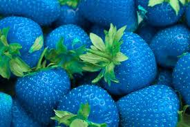 Deliciosos morangos com gosto e cor de revolver de feira. Somente tecnologia GMO pode fazer essa delicia.
Sem GMOs o mundo nao conseguira produzir o tanto de alimento que vai consumir. GMOs podem ajudar tanto no combate a pragas quanto no muito melhor aproveitamento da energia solar e agua, aumentando em muito a produtividade da fotossintese, ao passo que os alimentos organicos puros de origem nao produzem muita coisa por unidade de terra (ainda que se bem cultivados fazem com que o solo volte a ser vivo, seguram erosao, promovem vida fora e dentro da biosfera).
Como ex-agronomo que acompanha o mercado vos digo que nao temos 'outra alternativa' (SIC); Nao ha tecnologia em vista para se aumentar a producao que nao seja atraves de GMOs. Obvio que ha riscos para fauna/flora nativa dos locais nos casos de GMOs que carregam inseticidas e genes resistentes a herbicidas, mas tambem ha coisas muito uteis como aumento de vitaminas e minerais dentro das plantas (ainda que ''artificialmente embutidos''). Exemplo? Ferro a mais em arroz plantado na Asia. Ferro combate -- entre outras coisas-- anemia. E black blocs. GMOs podem ser utilizados tambem positivamente aos humanos, ao contrario que os ''ativistas ecologicos'' pregam pelo planeta.
Alimento organico deveria ter prioridade em lugares onde a producao de graos principalmente nao seja possivel. Que tal terrenos vazios que tanto existem em todas as cidades? Ou aqueles locais onde maquinario nao pode operar por conta de declividade ou mesmo custos? Prefeituras e governos poderiam alugar terrenos que estao ociosos. Sem superfaturamento de alugueis e arrendamentos.
Wednesday, December 18, 2013
Terroir, Fim Do Misterio?
Um dos grandes mitos da viticultura parece que ja era. Lo siento muchachos, mas para tudo ha uma explicacao, ate para o "misterioso" conceito de terroir....
E ainda ha quem pense que americano nao sabe fazer vinhos, alias, ha quem escreva dizendo isso. Sou O fan de vinho europeu, mas quem sabe o que e como se faz esta nos EEUU. No caso americano laboratorio tambem he usado para o bem...ao contrario de uma tchurma que usa alquimia mesmo (novo mundo em geral, mas nao totalmente)
Ainda bem que nao fui estudar em Davis, passaria vergonha por la. Boa leitura, adieu terroir cabeca de bacalhau.
Link abaixo.
http://extra.globo.com/noticias/saude-e-ciencia/dna-revela-que-micro-organismos-sao-essenciais-para-sabor-do-vinho-10892936.html
 A verdadeira french connection....
E ainda ha quem pense que americano nao sabe fazer vinhos, alias, ha quem escreva dizendo isso. Sou O fan de vinho europeu, mas quem sabe o que e como se faz esta nos EEUU. No caso americano laboratorio tambem he usado para o bem...ao contrario de uma tchurma que usa alquimia mesmo (novo mundo em geral, mas nao totalmente)
Link abaixo.
http://extra.globo.com/noticias/saude-e-ciencia/dna-revela-que-micro-organismos-sao-essenciais-para-sabor-do-vinho-10892936.html
A verdadeira french connection....
Tuesday, December 17, 2013
Dica Cultural
Saudavel obsessao: Saber mais sobre o
passado para entender o presente e prever o que vem por ai. Cliche necessario de
lado, vai aqui mais um livro that fits the bill, nesse caso o bem escrito ‘a
ditatura derrotada’, de Elio Gaspari.
Livro gira em torno de Geisel e Golbery.
Duas figuras que se apresentam na minha memoria infantil apenas.
De espantoso sobre o livro? Que a historia
no Brasil se repete em intervalos muito mais curtos que parece e que continuamos
na mesma estrada do tal futuro espetacular e imediato que nunca chega.
Em 1970
e poucos o povao no RJ ja destruia trens em protestos, a economia era digna de
elogios do American Treasury Secretary, eramos o modelo a ser seguido. E a
bolsa de valores apresentava aumentos insustentaveis. Meu pai que o diga, levou
um p tombo.
Nao sou pro-ditadura, a nao ser que eu seja o ditador, mas da
nojo de comparar o patrimonio de vida de alguem como Geisel (ou ate mesmo o Figueiredo,
que figura) com os politicos/presidentes de hoje.
Mais um livro lido e aprovado pelo JRatao
Institute of Vague and Superficial, Albeit Necessary Culture, LLC. Livro doado por minhas secretarias loiras gemeas, a quem agradeco de publico.
Monday, December 16, 2013
Pequenos Vinhos, Grandes Preços
No champagne, no vinho argentino, chileno, americano, no brunello, no barolo ha os vinhos do caipira que quer se mostrar ou daqueles que estão simplesmente aprendendo. Cometemos erros quando somos iniciantes no vinho e geralmente os erros diminuem a medida que o tempo passa e a cintura fica mais aparente.
Tomei duas safras de Barolo Pio Cesare na vida. Sabado passado tomei a 2008. Um vinho pobre pelo preço. Muito so-so por tanto que se fala por ai. Mas quem tanto fala dele por ai? Juvenis, claro. Ninguem que ja tomou um bom barolo na vida classificaria esse tiozinho entre os grandes.
Barolo, Brunello e Champagne sao grandes decepcoes para muito enofilo iniciante por ai. No reveillon abrem champagnes de R$ 300.00 e na hora da celebração ninguém tem coragem de dizer que o vinho nao é grande coisa.
É como ir a um velório e dizer que o sanduíche (caro) estava ruim. Deixa para la.
Cuidado com vinhos que são famosos nas bocas e canetas de muitos.
Se beber, nao blogueie.
Possivel Catastrofe Em Marcha Na Europa?
O maior risco sempre fica com o produtor esse heroi desvalorizado. Quem ja plantou sabe como he.
http://www.oliveoiltimes.com/olive-oil-basics/fears-disease-could-spell-disaster-for-europes-olive-trees/36898
http://www.oliveoiltimes.com/olive-oil-basics/fears-disease-could-spell-disaster-for-europes-olive-trees/36898
Saturday, December 14, 2013
Leitura De Sabado.
Homens x mulheres. Diferentes, sim, melhores talvez....
Vive la différence!A new technique has drawn wiring diagrams of the brains of the two sexes. The contrast between them is illuminating.
MEN and women do not think in the same ways. Few would disagree with that. And science has quantified some of those differences. Men, it is pretty well established, have better motor and spatial abilities than women, and more monomaniacal patterns of thought. Women have better memories, are more socially adept, and are better at dealing with several things at once. There is a lot of overlap, obviously. But on average these observations are true.
Suggesting why they are true in evolutionary terms is a game anyone can play. One obvious idea is that because, in the days of hunting and gathering, men spent more time wandering away from camp, their brains needed to be adapted to able to find their way around. They also spent more time tracking, fighting and killing things, be they animals or intrusive neighbours. Women by contrast, politicked among themselves and brought up the children, so they needed to be adapted to enable them to manipulate each other’s and their children’s emotions to succeed in their world.
Finding out why sex differences are true in neurological terms—in other words, how the brain is wired up to create them—is another matter altogether. To play this game you have to have a lot of expensive kit, not just a comfortable chair from which to pontificate. And that is exactly what Ragini Verma of the University of Pennsylvania and her colleagues do have. As a result, as they outline in the Proceedings of the National Academy of Sciences, they have been able to map out differences in the ways male and female brains are cabled and match them, at least to their own satisfaction, to the stereotypes beloved of both folklore and psychology.
Sugar and spice or puppy-dogs’ tails?
Neurology has been revolutionised over the past couple of decades by a range of techniques that can scan living brains. Dr Verma’s technique of choice is diffusion tensor imaging. This follows water molecules around the brain. Because the fibres that connect nerve cells have fatty sheaths, the water in them can diffuse only along a fibre, not through the sheath. So, diffusion tensor imaging is able to detect bundles of such fibres, and see where they are going.
Dr Verma and her team applied the technique to 428 men and boys, and 521 women and girls. Their results are summarised in the two diagrams above, which show connection trends averaged from the sum of participants’ brains.
The two main parts of a human brain are the cerebrum, above and towards the front, which does the thinking, and the cerebellum, below and towards the back, which does the acting. Each is divided into right and left hemispheres. As the diagrams show, in men (the left-hand picture) the dominant connections in the cerebrum are those, marked in blue, within hemispheres. In women, they are those, marked in orange, between hemispheres. In the cerebellum (not visible because it is under the cerebrum), it is the other way around.
What this means is open to interpretation, but Dr Verma’s take is that the wiring differences underlie some of the variations in male and female cognitive skills. The left and right sides of the cerebrum, in particular, are believed to be specialised for logical and intuitive thought respectively. In her view, the cross-talk between them in women, suggested by the wiring diagrams, helps explain their better memories, social adeptness and ability to multitask, all of which benefit from the hemispheres collaborating. In men, by contrast, within-hemisphere links let them focus on things that do not need complex inputs from both hemispheres. Hence the monomania.
When it comes to the cerebellum, the extra cross-links between hemispheres in men serve to co-ordinate the activity of the whole sub-organ. That is important because each half controls, by itself, only one half of the body. Hence men have better motor abilities—or, in layman’s terms, are better co-ordinated than women.
Dr Verma’s other main finding is that most of these differences are not congenital. Rather, they develop with age. Her volunteers ranged from 8 to 22 years old. The brains of boys and girls aged 8 to 13 demonstrated only a few differences, though all were of the type that later became pronounced. Adolescents, those aged 13 to 17, showed more. Young adults, over 17, more still. Sex differences in brains—those visible to this technique, at least—thus manifest themselves mainly when sex itself begins to matter.
Dr Verma’s work is important not only for what it has shown, but also as a demonstration of the power of diffusion tensor imaging. Studying the brain, particularly the living brain, is a uniquely hard scientific problem. The American government has recently promised to spend serious amounts of money doing so, through what it dubs the BRAIN initiative—the inevitable contrived acronym supposedly standing for Brain Research through Advancing Innovative Neurotechnologies.
As that name suggests, advances in brain research depend on the development of better ways of looking at brains while they are alive and firing. This will be hard. But work like Dr Verma’s shows the rewards of doing it.
Friday, December 13, 2013
Feliz Imposto Velho No Natal e Fim De Ano
Nesse natal e fim de ano (segundo calendario cristao) lembre-se que voce pagara mais ou menos em torno de 80% de impostos nos espumantes.
Pensar que eu ria dos amigos escandinavos quando diziam que havia muito imposto embutido nos precos das bebidas com alcool por la....
Sei do que escrevo porque pago essa coisa. E melhor ainda: O uso que fazem dos impostos. Assunto velho? Sim. Tem solucao? Nao aqui.
Melhor fazer alcool com pao embolorado e chupar o negocio....
Thursday, December 12, 2013
Mais Uma Mafia Do ICMS Pega. Agora Voce Sabe De Onde Vem Milagres?
Deu no claudicante financeiramente falando OESP:
Fraudadores em varios estados de ICMS e ST foram pegos, alguns ja estao foragidos, etc etc. R$ 150,000,000 em evasao, aproximadamente.
Nao tem milagre no mundo financeiro, do comercio: Visivelmente alguns lojistas e distribuidores lesam o fisco e o mercado com os precos que praticam com constancia. Nao tem como vender um produto abaixo do preco do produtor ou importador se nao for contrabando, roubado, evasao fiscal (contrabando serve como evasao tambem), desespero, dumping ou compra de marketshare (expressao meio em desuso).
Em SP, Capital, ha gente que faz milagres ha muito tempo. As vezes pagam DARFs e ICMS PESADOS como multa. E voce achando que o velhinho da distribuidora era honesto por praticar precos tao mais baixos que outrem.
Nao ha milagre. Ha gente que abusa dos precos e os larapios que em nome de riqueza facil/rapida estao enquadrilhados.
Acorda Ze Ruela. Nao compre nada pirata nem produto roubado/contrabandeado.
Wednesday, December 11, 2013
Melhores De SP, Veja Edicao Fim De Ano
Boas noticias, e mais do de sempre.
Primeiro a turma que nao se emenda:
Ainda consta muita gente que vende espuma no prato por R$ 90.00, pedacos de arte.... tudo como era no Adria ha 10 anos. Enfiem essa espuma na pia e facam comida.
Quem nao pegou, pegue a nova Veja Melhores 2013 2014. Da para acompanhar a evolucao do mercado. Numero enorme de restaurante tradicional e famoso que fechou as portas. Paginas e paginas de gelaterias (antes eram 4, 5 das boas por toda SP), muita chocolateria (algumas mais caras que uma refeicao, mas sao otimas), mais cafes em pinheiros/vila madalena que mcdonald's em SP toda. Agora hamburguer virou coisa de rico... e me parece que as pessoas estao comecando a ensaiar uma rebeliao contra pizza cara. Por R$ 120.00 vamos pegar um espanhol/portuga/japa/italiano e ainda vem troco
De bom na parte de restaurantes? O surgimento com mais forza da liga dos bons e baratos: Que seja uma tendencia com muito futuro. E que os donos e investidores tenham sucesso tambem.
Mais uma coisa: Os japas que eram muito caros ha 8 anos continuam com o mesmo preco. Realmente nao daria para subir os precos no mesmo passo que a inflacao. Entao hoje um otimo japa por R$ 260.00 no menu confianza esta bem mais palatavel que antes.
Recomendo o kinoshita e o sakamoto. Uma vez por ano da para pagar R$ 300.00 nesses lugares. Valem o custo. Eu ainda prefiro o kinoshita.
Monday, December 9, 2013
Grandes Mentiras Do Vinho. Dialogos Epicos
Cada dia mais orgulhoso dos enopicaretas.
Da serie ''ainda nao vi tudo em lojas de vinhos'':
P: De que safra eh esse vinho branco?
Resposta do cara de pau: Esse otimo vinho he um blend de safras. Todas muito boas.
P: Mas o vinho esta novo?
Resposta: Esta otimo, pode leva-lo.
-------------------------------------------------------------------------------------------------------
Blend de safras? O ''vinho'' em questao deve ser um blend de aldeidos, compostos inorganicos e metais radioativos.
Saturday, December 7, 2013
Leitura De Sabado:
Religiao faz o louco ou o louco cria religiao porque he doido?
http://www.theguardian.com/uk-news/2013/dec/06/muslim-vigilantes-jailed-sharia-law-attacks-london
http://www.theguardian.com/uk-news/2013/dec/06/muslim-vigilantes-jailed-sharia-law-attacks-london
Friday, December 6, 2013
Fim Do Vinho Ruim?
Um grande artigo, para ler com gosto. Pergunta: vinho natural ruim, mas feito pela natureza ou vinho "bom" feito por computador, maquinas??
http://www.economist.com/news/technology-quarterly/21590767-high-tech-winemaking-technology-has-already-made-poor-plonk-thing-past
http://www.economist.com/news/technology-quarterly/21590767-high-tech-winemaking-technology-has-already-made-poor-plonk-thing-past
Thursday, December 5, 2013
Pequenos Vinhos, Grandes Precos
Ay ay ay. Houve uma epoca que eu pensava que esse vinho fosse um dos melhores vinhos do Chile ainda que nao tivesse jamais degustado essa coisa. Somente ouvia dizer que '' ''.
Nao faz muito vi algumas garrafas no caixa do supermercado (pasme) Dia.
E acabei morrendo em R$ 90.00 por uma garrafa de Marques de Casa Concha Cabernet Sauvignon 2008. Uma mega decepcao pelo preco e pela suposta historia que tem no Brasil. Segundo uma pesquisa de mercado rapida o preco ate que nao estava ruim.
O que? Voce quer ficar multi-bilionario cobrando precos justos pelos seus produtos? Dream on, babe.
Perguntar Ofende: Voce Sabe Definir Com Exatidao O Que He Um Produto Organico?
Aposto que nao. No mundo todo os paises tem definicoes na lei diferentes para os produtos chamados de organico. Porcentagens variam, agrotoxico para ca e para la tambem.
E essa da semente (ler abaixo). Problema? Nenhum desde que as dosagens e doses sejam respeitadas de acordo com o que manda o fabricante....mas dai o produto ja nao he mais ''organico'' 100%....
http://www1.folha.uol.com.br/mercado/2013/12/1381025-alimentos-organicos-continuarao-a-poder-ser-produzidos-com-sementes-com-agrotoxicos.shtml
 I'm 100% carbon based life form...therefore organic!!
E essa da semente (ler abaixo). Problema? Nenhum desde que as dosagens e doses sejam respeitadas de acordo com o que manda o fabricante....mas dai o produto ja nao he mais ''organico'' 100%....
http://www1.folha.uol.com.br/mercado/2013/12/1381025-alimentos-organicos-continuarao-a-poder-ser-produzidos-com-sementes-com-agrotoxicos.shtml
Tuesday, December 3, 2013
A Triste Historia Dos Azeites De Oliva Fraudados Continua
So ler aqui. E voce achando que esta colocando saude no seu prato....e da sua familia
http://www.procon.rj.gov.br/index.php/publicacao/detalhar/775
http://www.procon.rj.gov.br/index.php/publicacao/detalhar/775
Monday, December 2, 2013
A Importancia Do Trouxa Para O Comercio
Once upon a time eu morei 365 dias exatos numa cidade aprazivel no Bay area. Essa cidade tem uma ponte que he a mais linda do mundo. O nome da cidade fica incognito aqui. Mas foi o melhor ano da minha vida.
Durante os 365 dias havia no Market Street lojas com faixas dizendo ''fechando as portas, liquidacao total''. Blowout sale, going out of business.
Market St he passagem obrigatoria para moradores e muitos turistas. Atraidos pela oportunidade unica na vida de comprar coisas em SFO por preco de ocasiao, milhares de pessoas passavam por ali e deixavam a carteira na compra de coisas inuteis por precos que nao eram exatamente a blowout.
Defendo aqui que o tonto, o trouxa, o comprador sem logica, impulsivo (mulheres e homens) sao muito importantes para o comercio, comunidades locais, governo, etc.
Se todos fossem como eu (racional e conservador na compra, apesar de investir pesado em negocios em relacao ao patrimonio, mas isso he outra coisa) ou se fossem como varios clientes que tenho que so compram galinha morta, vinhos ou produtos REALMENTE com precos muitos bons, estariamos ainda produzindo carros como Ford T preto, somente para a zelite.
Esse cara que eu chamo de enotrouxa cria (transfere) riquezas. Ao comprar um vinho que custou na origem R$ 18.00 (preco nacionalizado, com impostos incluidos) por R$ 120 o cliente esta movendo e acelerando a roda do comercio, eventualmente abrindo empregos e fazendo com que mais impostos vao ao governo.
Nao haveria comercio de imoveis atualmente se nao houvessse o trouxa (R$ 15,000 o m2??)
No caso do vinho, para ser mais especifico eu chuto aqui que ha dois tipos de trouxas: Aquele contumaz que jamais vai aprender que aquele ''champagne'' feito no RGS que ele compra por R$ 70.00 he realmente um lixo. Esse nunca vai aprender. Esse vai comprar calca rasgada de R$ 1,200 nas boutiques da Oscar Freire achando que fez um puta negocio.
Mas tambem temos o trouxa passageiro. Pessoas como voce e eu que ja pagaram caro em vinhos asquerosos, mas com um pouco de tempo fomos lendo, conversando e aprendendo sobre a loucura que cometiamos. Hoje somos racionais e chatos para comprar.
Esse cidadao rotulado carinhosamente de ''trouxa'' merece nossa atencao, respeito e admiracao. Obrigado por transferirem a economia muito mais que deveriam.
Subscribe to:
Posts (Atom)